Image of Whitlow Au and the dolphin BJ.

Whitlow Au awarded gold medal by Acoustical Society of America

Whitlow W. L. Au, emeritus research professor at the Hawaiʻi Institute of Marine Biology (HIMB), has been named recipient of the gold medal of the Acoustical Society of America (ASA) for contributions to understanding underwater biosonar and for service to the acoustical society. The gold medal, which will be presented at the 171st meeting of the ASA on May 25, is given to a member of the ASA whose contributions to the field of acoustics and to the acoustical society have been unusually distinguished.

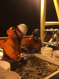
Au’s research has been devoted to understanding dolphin biosonar and to learning why it is so superior to man-made sonar. This involves understanding the properties and capabilities of dolphin biosonar, discovering how dolphins produce sound and how sound travels through the head and enters the water via their foreheads. It also involves understanding how the “special” type of signals produced by dolphins capture the essence of different objects. The ultimate goal is to design sonars that are based on the principles underlying dolphin biosonar.

“I am honored and humbled to receive this award. This is the first time that the Gold Medal has been awarded to someone who performed most of his research in Hawaiʻi,” said Au.

Au was born and raised in Hawaiʻi and attended St. Louis High School. He earned a bachelor’s of science in electrical engineering from the University of Hawaiʻi and a doctoral degree in electrical sciences from Washington State University. He served as a senior scientist at the Naval Ocean Systems Center, Hawaiʻi Laboratory from 1971 to 1993 after which he joined the Marine Mammal Research Program at HIMB as chief scientist where he continues today as emeritus research professor.

Sonar was invented just prior to World War I. However, dolphins have been using “biosonar” for millions of years to search for food and avoid obstacles. In shallow water and for short ranges below 200–300 meters evolutionary pressures over millennia have resulted in dolphin biosonar being considerably superior to man-made sonar.

“Doing research in science is fun and I am blessed to be able to spend a career having fun,” said Au.

Contributions to the scientific community

Au served on the National Research Council Ocean Studies Board (2004–06) and as chair or member of organizing committees for national and international meetings and symposiums including the International Symposium on Sensory Systems and Behavior of Aquatic Mammals (1991), the 3rd International Symposium on Animal Sonar (1996), the Acoustic Communication by Animals International Symposium series (2008, 2010, 2011), and Joint Meetings of the ASA and the Acoustical Society of Japan in 1996, 2006, and upcoming in the fall of 2016.

His service to ASA includes Chair of the Technical Committee on Animal Bioacoustics (1997–00), associate editor, Journal of the Acoustical Society of America for Animal Bioacoustics (1998–), member of the executive council (2001–04), vice president (2006–07) and president 2009–10). He was awarded the first ASA silver medal in animal bioacoustics in 1998.

Image of fish and coral reef in PMNM

Protecting diversity on coral reefs: DNA may hold the key

Coral reefs are widely known for their stunning array of color, shape and forms of life, making them a model for extreme biodiversity. Hidden within the multitude of reef inhabitants, but no less important, is their genetic diversity— variability in DNA that gives species the capacity for adaptation, speciation and resilience in the face of stress.

Research published on April 27 by a team of scientists from the University of Hawaiʻi at Mānoa School of Ocean and Earth Science and Technology, the University of California, Santa Barbara (UCSB), University of St. Andrews and University of Melbourne discovered that large areas of intact coral reef with extensive live coral cover, not disturbed by humans or climate change, harbor the greatest amount of genetic diversity.

The team, led by Kimberly Selkoe, associate researcher at the UH Mānoa Hawaiʻi Institute of Marine Biology(HIMB) and UCSB, assessed genetic diversity from more than 17,000 samples taken from 47 common reef-associated species across the Hawaiian Archipelago. With this work, the researchers uncovered a link between species diversity of an ecosystem and the genetic diversity encoded within the DNA of those species.

New information directs conservation efforts

Genetic diversity is linked to adaptive capacity, extinction risk of species and the overall functioning of ecosystems, so conservation strategies often call for preserving areas of high genetic diversity. However, prior to this study there was little direct data to guide debates such as whether many small versus one large marine reserve would be better to protect genetic resources. The study found that Papahānaumokuākea Marine National Monument safeguards the majority of Hawaiian reef biodiversity, but populations in the main Hawaiian Islands (MHI) are genetically distinct and warrant additional protection.

“Management of marine resources should be based on the best available science, and this study underscores the importance of protecting large areas of coral reef habitat,” said Randall Kosaki, NOAA’s deputy superintendent of Papahānaumokuākea. “Protecting biodiversity and ecosystem integrity is our primary goal.”

“Our study provides support for campaigns such as the Big Ocean Initiative, that seek to protect large areas of healthy coral reef habitat,” said Rob Toonen, research professor at HIMB and co-author of the study. “However, we also need a diversity of approaches to preserve marine biodiversity because one size simply does not fit all in conservation efforts.”

Unique features in the main and northwestern Hawaiian Islands

The researchers found that genetic diversity varies across the inhabited main Hawaiian Islands, extending the argument for targeted protection of reefs throughout the main Hawaiian Islands. Notably, Hawaiʻi Island has the greatest amount of coral reef area, harbors the greatest proportion of genetic diversity, and serves a unique, and particularly influential role due to its large size at the margin of the chain. Nihoa and Niʻihau, which sit at the transition between Papahānaumokuākea Marine National Monument and the main Hawaiian Islands, also stand out and warrant future research and protection due to intriguing combinations of high genetic divergence and unusual fish composition.

Capacity for adapting to climate change

Genetic diversity is widely accepted as critical for adaptation to a changing climate. Although coral bleaching in Hawaiʻi has been relatively rare over the past few decades by comparison to many other places in the Pacific, this stressor still exerts a large impact on the reef communities studied. Researchers found that areas which experienced the most frequent coral bleaching conditions also showed depressed levels of genetic diversity, not just for corals but for the entire community of fishes and other invertebrates associated with those habitats.

“This negative impact of thermal stress on genetic diversity suggests that climate change will compromise the adaptive capacity and genetic integrity of not just corals but the entire coral reef community,” explained Selkoe.

Identifying features of the landscape that are associated with biodiversity hotspots is one of the key strategies for conservation. In the future, the team will follow up on intriguing evidence in the dataset that the same large-scale ecological rules that predict species diversity also predict genetic diversity.

“If these relationships can be confirmed and replicated elsewhere, DNA sampling may one day allow rapid assessment of species diversity and new metrics of resilience and adaptive capacity,” explained Selkoe, also an associate at UCSB’s National Center for Ecological Analysis and Synthesis and an adjunct professor at the campus’ Bren School of Ecological Science and Management.

Image of juvenile lace coral

Coral toolkit allows floating larvae to transform into reef skeletons

In a study published today, researchers from the University of Hawai‘i at MānoaRutgers University and the University of Haifa identified key and novel components of the molecular “toolkit” that allow corals to build their skeletons (called biomineralization) and described when—in the transformation from floating larvae to coral skeleton—these components are used.

Corals are the sum of a symbiotic relationship between cnidarian animals and millions of single-celled algae that live inside their tissues. These symbiotic algae photosynthesize and provide the energy corals need to build their skeletons. In turn, the skeletons form the structure and framework of coral reefs that provide habitat to fishes and other animals. Coral reef are so large they can be seen from space, and generate goods and services valued on the order of billions of dollars annually.

Natural laboratory

The reefs of Kāneʻohe Bay, Hawaiʻi around the Hawaiʻi Institute of Marine Biology (HIMB) at the UH Mānoa School of Ocean and Earth Science and Technology provided the ideal natural coral study system.

“Our research on reproduction in the lace coral, Pocillopora damicornis, provided the perfect opportunity to look at a natural on-off switch in coral biomineralization,” said co-lead author Hollie Putnam, HIMB assistant researcher.

Revealing the mechanics of transformation

Corals release their offspring as swimming larvae with no skeleton. The larvae change shape, settle onto the reef, and start to build their skeletons. Putnam and Ruth Gates, senior researcher and director of HIMB, collaborated with scientists from Rutgers University, and the University of Haifa to examine gene expression and the production of proteins at these different life stages.

“Together, we described components that were ‘off’ or ‘on’ before and after the first skeletal structures were built,” said Putnam. “This approach revealed the quantity and location of important components of the biomineralization machinery, knowledge that allows them to be developed as biomarkers for studies of coral growth in the future.”

Understanding growth leads to better management

Coral reefs are undergoing mass mortality due to high seawater temperatures and ocean acidification is diminishing their capacity for skeletal growth. These global stressors are superimposed on the local threats of coastal development, excess nutrients and overfishing; and all combine to threaten the persistence of corals as a functional ecosystem.

“The better we understand how corals grow, the better we understand the way they respond to ocean acidification, rising sea surface temperatures and pollution and can therefore forecast and manage reefs communities in the future,” said Gates.

The team is currently working to further describe these novel components of the biomineralization machine in corals and how they are regulated under increased environmental stress.

Coral reef with algae

Microbial takeover on coral reefs?

Coral reefs—the world’s most productive and diverse marine ecosystems—rely on a masterful recycling program to stay healthy. The corals and algae that form the base of the reef’s food web release a variety of nutrients that support a complex and efficient food chain. But when this system gets out of whack, the cycle breaks down and endangers the coral reef’s health. A new study led by researchers at San Diego State University (SDSU) and co-authored by assistant professor Craig Nelson at the University of Hawaiʻi at Mānoa (UHM) Center for Microbial Oceanography: Research and Education explores how a process called microbialization destroys links in this delicate food chain.

Millions of people around the world depend on coral reefs to provide productive fisheries, and they play an important role in global environmental health. Overfishing the waters near reefs, however, removes the primary consumers of algae, allowing fleshy algae to spread unchecked. In areas with large human populations, pollution often exacerbates the problem by stimulating the algae.

How more algae brings more microbes

Fleshy algae on reefs exude copious amounts of nutrients known as dissolved organic carbon (DOC), which microbes eat. Researchers theorized that when reef ecosystems have elevated levels of algae producing meals for microbes, higher levels of potentially harmful microbes can occur throughout the reef ecosystem. In this newly abundant population of microbes, evolutionary selection pressures favor microbes that endanger corals, either by depleting oxygen from the environment or through disease.

As the corals die off, the algae have even more space to take over, producing more DOC and creating a runaway feedback loop that leads to further coral mortality and microbes taking over the ecosystem, a process the scientists have termed “microbialization”.

“Reefs dominated by algae show a fundamental change in the way carbon and nutrients are recycled by microbes,” said Nelson.

“Most of the energy in the ecosystem goes into the microbes,” said the study’s lead author, Andreas F. Haas, a biologist at SDSU. “It doesn’t support the variety of reef organisms which make up a healthy system anymore.”

Testing the theory

Haas, Nelson and the international team of researchers set out to test this theory by collecting more than 400 water samples from 60 coral reef sites across the Indian, Pacific and Atlantic Oceans. In the laboratory, they tested these samples for evidence of microbialization of algae-dominated reefs worldwide: more microbes with more potential to harm reef organisms.

First, they analyzed the abundance of microbes throughout their samples. Supporting their hypotheses, they found that reef sites with higher algal cover also had more microbes. Using metagenomic sequencing techniques, they found that in algae-dominated reefs, the microbial community is more likely to harbor harmful pathogens.

This pattern has geochemical implications for the ocean carbon cycle, as well. One of the counterintuitive predictions made by the model is that the microbes fostered by algal growth are voracious, stripping the reef of DOC and limiting the transfer of organic material to larger organisms like invertebrates and fish. Sure enough, the team found that in reefs with high algal cover, such as the island of Kiritimati in the central Pacific Ocean, DOC concentrations were very low, whereas in reefs with low algal cover, such as near the Kingman reef in the north Pacific Ocean, DOC was higher. Across the 60 sampling sites and across three ocean basins they found this relationship held true: the higher the algal cover, the lower the DOC.

“Algae always release more dissolved organic carbon than corals,” Haas said, “but in the reefs with more algae you see less DOC.”

The big picture

In short, the study’s results support the idea that microbialization associated with increasing algae cover in coral reefs can decimate the reef ecosystem through microbial takeover. The researchers published their findings today in the journal Nature Microbiology.

As overfishing and eutrophication are two of the leading causes of increased algal cover, humans should be concerned about how their actions both directly and indirectly impact one of the world’s most important ecosystems, the researchers concluded.

* * *

Haas, A.F., M.F.M. Fairoz, L.W. Kelly, C.E. Nelson, Y.W. Lim, B. Knowles, E.A. Dinsdale, R.A. Edwards, S. Giles, M. Hatay, N. Hisakawa, H. Maughan, O. Pantos, T. Roach, S.E. Sanchez, S. Sandin, J.E. Smith and F. Rohwer. Global microbialization of coral reefs. In Press. Nature Microbiology. DOI: 10.1038/nmicrobiol.2016.42

See UH News for more images.

Research expedition blog online: climate change and Antarctic fjord ecosystem

Follow the research activities of scientists with the FjordEco project (from SOEST, Scripps Institution of Oceanography and the University of Alaska at Fairbanks) as they study fjord ecosystem dynamics in Antarctica.

Marine communities along the Western Antarctic Peninsula (WAP) are highly productive ecosystems which support a diverse assemblage of charismatic animals such as penguins, seals, and whales as well as commercial fisheries including for Antarctic krill. The WAP also contains many fjords (deep estuaries carved by glacial ice) with active glaciers entering the ocean;  these fjords appear to be intense, potentially climate sensitive, hotspots of biological production and biodiversity. Because of intense biological activity and abundant charismatic fauna, these fjords are also major destinations for a large Antarctic tourism industry. Nonetheless, the structure and dynamics of these fjord ecosystems are very poorly understood.

Craig Smith and graduate student Astrid Leitner of UH sorting animals from the Blake trawl in the wee hours of a cold night.

Craig Smith and graduate student Astrid Leitner of UH sorting animals from the Blake trawl in the wee hours of a cold night.

The FjordEco project is an integrated field and modeling program designed to evaluate physical oceanographic processes, glacial inputs, water column community dynamics, and seafloor bottom community structure and function in these important yet little understood fjord systems. These Antarctic fjords have characteristics that are substantially different from well-studied Arctic fjords, likely yielding much different responses to climate warming. FjordEco is designed to provide major new insights into the dynamics and climate sensitivity of Antarctic fjord ecosystems, highlighting contrasts with Arctic sub-polar fjords, and potentially transforming our understanding of the ecological role of fjords in the rapidly warming west Antarctic coastal marine landscape.

Heat trumps cold in the treatment of jellyfish stings

A recent study by researchers at the University of Hawai‘i – Mānoa (UHM), published this month in the journal Toxins, may finally put to rest the ongoing debate about whether to use cold or heat to treat jellyfish stings. Their systematic and critical review provides overwhelming evidence that clinical outcomes from all kinds of jellyfish stings are improved following treatment with hot packs or hot-water immersion.

Jellyfish stings are a growing public health concern worldwide and are responsible for more deaths than shark attacks each year. Even “mild” stings can hurt for hours to days and leave lasting scars. Despite the danger posed by these gelatinous invertebrates, scientists and medical professionals still do not agree on the best way to treat and manage jellyfish stings. At the center of the contentious debate is whether it is better to treat the sting site with heat or cold.

“People think ice will help because jelly stings burn and ice is cold,” said Dr. Christie Wilcox, a postdoctoral fellow at the John A. Burns School of Medicine (JABSOM) and lead author of the paper. “And if you Google it, many sites – even those considered reputable – will tell you to put ice on a sting to dull the pain. But research to date has shown that all marine venoms are highly heat sensitive, thus hot water or hot packs should be more effective than cold packs or ice.”

“Authoritative web articles are constantly bombarding the public with unvalidated and frankly bad advice for how to treat jelly stings,” said Dr. Angel Yanagihara, assistant research professor at the UHM Pacific Biosciences Research Center (PBRC) and JABSOM and senior author of the paper, who for the past 18 years, as Director of the Pacific Cnidaria Research Laboratory, has studied the pathophysiology of jellyfish stings. “In Hawaii, and around the world, we have seen that first responders and public health decision makers rely on non-evidence-based claims found on websites. It’s not too strong to point out that in some cases, ignorance can cost lives. We conducted this study to rigorously assemble all the published data in hopes that policy makers will revisit this issue and carefully consider the available evidence. We are also engaged in new experimental work with models looking at vinegar effects, as well as well-designed randomized clinical trials. The goal of my laboratory’s efforts is to contribute to evidence-based best clinical practices for jellyfish stings.”

Wilcox and Yanagihara conducted a systematic review to compare the use of cold or heat in jellyfish sting treatment using a common ranking system for clinical evidence. The pair combed through more than 2,000 related articles from searches of major scientific journal article databases to find every study to date that examined the effects of using temperature-based treatments for jellyfish stings. The overwhelming preponderance of evidence supported the use of hot-water immersion (about 45 degrees Celsius). This is consistent with findings in more than a dozen articles, demonstrating that venom components are inactivated at temperatures between 40 and 50 degrees Celsius.

“I was shocked that the science was so clear, given that there is so much debate over the use of hot water,” said Wilcox. Hot-water immersion is already the standard of care for other severe marine envenomations including potentially life-threatening stonefish stings, so these results help streamline the first-aid response. “It’s simple, really: if you’re stung, use hot water or hot packs rather than ice or cold packs.”

Image from µCT scan highlighting bioerosion in a coral block

New imaging technique reveals vulnerability of coral reefs

Corals, the primary reef builders on coral reefs, are often the star player in research studies addressing the impacts of climate change on coral reefs because they are the foundation of coral reef ecosystems. However, the breakdown of coral reefs from borers (such as bivalves, sponges, and marine worms) and grazers (such as parrotfish and urchins) – called bioerosion – and growth from encrusting algae and invertebrates (for example, oysters and barnacles) – called secondary accretion – are critical processes for reef sustainability.

In a study published today in PLOS ONE, Nyssa Silbiger and colleagues created a novel method to expose how these underdogs of coral reef science respond to varying environmental conditions, including changing ocean acidity.  Using µCT (micro-computed tomography) scans, Silbiger and colleagues were able to calculate detailed bioerosion and secondary accretion rates on coral reefs – work she performed as a graduate student at the University of Hawai‘i – Mānoa’s (UHM) Hawai‘i Institute of Marine Biology (HIMB).

New application of proven technology

Silbiger, now a post-doctoral scholar at the University of California – Irvine, and colleagues used fine-scale µCT scans – a technique that is commonly used in the medical field to image internal organs and bones in 3D – to create 3-dimentional images inside and outside of calcium carbonate blocks constructed from dead pieces of the coral skeleton. Silbiger and colleagues applied this non-destructive µCT technology to reveal the impacts of changing climate conditions on reef bioerosion and secondary accretion.

“While there are several methods currently being used to test how bioerosion responds to environmental variability, our study provides the first method to accurately separate bioerosion and secondary accretion on the same time-scale and determine how these processes individually respond to different environmental parameters,” said Silbiger.

Detailed scans provide new insight

The team of researchers constructed blocks from dead pieces of coral and performed µCT scans on the samples before and after a one-year deployment period on a coral reef in Kāne`ohe Bay, Hawai`i to determine how much new growth had settled onto the block and how much of the block had been eroded from bioeroding organisms. By comparing pre- and post-deployment µCT scans, Silbiger and colleagues were able to separate secondary accretion and bioerosion from the same experimental substrate exposed to the same environmental variation over the same time-scale. Because the blocks were placed along an environmental gradient with naturally varying pH conditions (acidity), the researchers could assess how pH and other environmental parameters impact secondary accretion and bioerosion.

In a previously published dataset (Silbiger et al. 2014 Marine Ecology Progress Series), Silbiger and colleagues showed that reefs shifted from net growth to net erosion along this natural pH gradient. However, with this new analysis the scientists were able to uncover results that were unattainable with prior methods. They demonstrated that secondary accretion and bioerosion are driven by different environmental parameters – bioerosion is more sensitive to changing ocean pH than secondary accretion, and the net change in reef growth is driven more by changes in bioerosion than secondary accretion.

“We were surprised that bioerosion was so much more sensitive to ocean acidification than growth processes on the reef,” said Silbiger, “This key finding could change our perspective on how coral reefs are predicted to respond to ocean acidification.”

Startling results and the promise of new discoveries

In order for coral reefs to persist, the rate of reef growth must be higher than the rate of reef breakdown. If reef breakdown continues to increase because of ocean acidification there could be a deadly shift from net growth to net breakdown of coral reefs in the future.

“The results of our study are sobering because it seems that even if corals can adapt, acclimatize or withstand changing ocean pH, bioerosion of the reef framework will still continue to increase,” according to Silbiger.

In order to predict how reefs will change in the future, it is critical to understand how environmental variability impacts individual reef processes. Without this information, researchers could over- or under-estimate how coral reefs will actually respond to future ocean conditions. Silbiger and colleagues are excited by the discoveries that await using µCT technology.

We are able to assess the addition or removal of calcium carbonate (the skeletal make-up of coral reefs) at a resolution of 100µm [micrometers] – approximately the thickness of a human hair,” said Silbiger. “There is so much that we can learn about coral reefs using µCT scans. My colleagues and I are mining all the information we can from this exciting technology.”

* * *

Silbiger NJ, Guadayol Ó, Thomas FIM, Donahue MJ (2016) A novel μCT analysis reveals different responses of bioerosion and secondary accretion to environmental variability. PLOS ONE: doi: 10.1371/journal.pone.0153058

All the µCT scans for this project were done by the Cornell University Multiscale CT Facility for Imaging and Preclinical Research.

Photo of Katie Barott filtering water

Piggybacking viruses

In the microscopic life that thrives around coral reefs, a team of researchers, including Katie Barott, postdoctoral researcher at the University of Hawai‘i – Mānoa’s Hawai‘i Institute of Marine Biology (HIMB), have discovered an interplay between viruses and microbes that defies conventional wisdom. As the density of microbes rises in an ecosystem, the number of viruses infecting those microbes rises with it. It has generally been assumed that this growing population of viruses, in turn, kills more and more microbes, keeping the microbial population in check. It’s a model known as “kill-the-winner”—the winners being the blooming microbial cells and the killers being the viruses (mostly bacteria-killing viruses known as bacteriophages) that infect them.

However, previous research has suggested that, under certain conditions, viruses can change their infection strategy. As potential host microbes become more numerous, some viruses forego rapid replication and opt instead to reside peaceably inside their host, thereby reducing the viruses’ numbers. In a study published recently in the journal Nature, Barott, along with lead author Ben Knowles, a viral ecologist at San Diego State University, and co-authors refer to this alternative model as “piggyback-the-winner,” and it could have implications for phage-based medicine and ecosystem resilience in the face of environmental disturbances that promote microbial blooms.

Microbial population explosions can take many forms—algal blooms in the ocean and in lakes, fungal blights in soil and bacterial infection in humans are just a few examples—and how viruses respond to this rapid microbial growth has long interested ecologists. Many viruses can make the switch between rapid replication and dormant coexistence. For decades, most researchers have assumed that during microbial population booms, their viruses take advantage of the opportunity to multiply by killing the abundant microbial winners.

“Kill-the-winner seems to make sense,” said Knowles. “The logic behind it has been around for a while. The reasoning is very seductive.”

And the winner is…

The international team of collaborators – with expertise ranging from mathematics, physics and statistics to ecology and molecular biology – decided to put this model to the test. They collected samples of microbe-rich seawater near coral reefs in both the Pacific and Atlantic Oceans. Then, using a combination of microscopic and genomic techniques, they analyzed those samples for the abundance and nature of both microbes and the viruses that infect them.

Under the kill-the-winner model, researchers would expect to find more viruses per microbe in samples with a high microbial density and growth rates. What the team found, however, was just the opposite: As microbial abundance increased, the virus-to-microbe ratio decreased significantly.

Next, the team ran an experiment in which they incubated seawater from a pristine coral reef location and from Mission Bay in San Diego for several days, during which they monitored the viral and microbial abundance. The results matched their field sampling, with virus numbers staying relatively low even as microbial populations bloomed.

Why weren’t the viruses exploiting the increasing population of hosts by infecting them and multiplying rapidly? Why weren’t they killing the winner? Exploring this phenomenon further, the researchers used metagenomic analysis to determine whether the viruses in the sample showed virulent, predatory traits or the hallmarks of non-predatory lifestyles. Intriguingly, they found that in samples with a higher microbe count, viral communities became less virulent.

Better living through integration

Instead of multiplying and killing off their booming host population, more of the viruses instead integrate themselves into their host. The viruses replicate more slowly, but they also avoid competing with other viruses and having to navigate with the host’s own immunity defenses. This piggyback-the-winner model better explains virus-host dynamics during periods of fast microbial growth than the established kill-the-winner model, the researchers said.

“When you have a fast-growing host, if you’re a virus, you profit more from integration,” Knowles said. “It’s just intelligent parasitism.”

A better understanding of these dynamics holds promise for improving human health. For example, specially targeted phages have been suggested as a possible therapy for conditions like cystic fibrosis, which is caused by frequent bacterial lung infections. This discovery also could help improve marine ecologists’ understanding of the microbiological forces that influence coral reef health.

Deep-sea sediment cores

Human carbon release rate is unprecedented in the past 66 million years of Earth’s history

The earliest instrumental records of Earth’s climate, as measured by thermometers and other tools, start in the 1850s. To look further back in time, scientists investigate air bubbles trapped in ice cores, which expands the window to less than a million years. But to study Earth’s history over tens to hundreds of millions of years, researchers examine the chemical and biological signatures of deep sea sediment archives.

New research published today in Nature Geoscience by Richard Zeebe, professor at the University of Hawai‘i – Mānoa School of Ocean and Earth Science and Technology (SOEST), and colleagues looks at changes of Earth’s temperature and atmospheric carbon dioxide (CO2) since the end of the age of the dinosaurs. Their findings suggest humans are releasing carbon about 10 times faster than during any event in the past 66 million years.

The research team developed a new approach and was able to determine the duration of the onset of an important past climate event, the Paleocene-Eocene Thermal Maximum, PETM for short, 56 million years ago.

“As far as we know, the PETM has the largest carbon release during the past 66 million years,” said Zeebe.

No good comparison

Zeebe and co-authors Andy Ridgwell (University of Bristol/ University of California) and James Zachos (University of California) combined analyses of chemical properties of PETM sediment cores with numerical simulations of Earth’s climate and carbon cycle. Their new method allows them to extract rates of change from a sediment record without the need for an actual sediment age model. Applied to the PETM, they calculated how fast the carbon was released, how fast Earth’s surface warmed, and constrained the time scale of the onset, which was at least 4,000 years.

The rate of carbon release during the PETM was determined to be much smaller than the current input of carbon to the atmosphere from human activities. Carbon release rates from human sources reached a record high in 2014 of about 37 billion metric tons of CO2. The researchers estimated the maximum sustained carbon release rate during the PETM had to be less than 4 billion metric tons of CO2 per year – about one-tenth the current rate.

“Because our carbon release rate is unprecedented over such a long time period in Earth’s history, it also means that we have effectively entered a ‘no-analogue’ state. This represents a big challenge for projecting future climate changes because we have no good comparison from the past,” said Zeebe.

Uncharted territory

Whereas large climate transitions in the past may have been relatively smooth, there is no guarantee for the future. The climate system is non-linear, which means its response to a forcing (such as our CO2 emissions) is a complex process involving a whole suite of components.

“If you kick a system very fast, it usually responds differently than if you nudge it slowly but steadily”, said Zeebe. “Also, it is rather likely that future disruptions of ecosystems will exceed the relatively limited extinctions observed at the PETM,” Zeebe added.

“In studying one of the most dramatic episodes of global change since the end of the age of the dinosaurs, these scientists show that we are currently in uncharted territory in the rate carbon is being released into the atmosphere and oceans,” says Candace Major, program director in the National Science Foundation (NSF)’s Division of Ocean Sciences, which funded the research.

The long view

Scientists like Zeebe also study the PETM to better understand long-term changes in Earth’s future climate. Most of the current climate debate concentrates only on this century but the PETM suggests that the consequences of our massive fossil fuel burning will have a much, much longer tail.

“Everyone is focused on what happens by 2100. But that’s only two generations from today. It’s like: If the world ends in 2100 we’re probably OK!” said Zeebe. “But it’s very clear that over a longer timescale there will be much bigger changes.”

Zeebe and his colleagues continue their work on the PETM to study other aspects of the event – for example, determining how severe ocean acidification was during the PETM and what impact it had on calcifying organisms in the ocean. This may provide insight about what to expect in the future as Earth’s climate continues to warm and oceans keep acidifying.

* * *

Richard E. Zeebe, Andy Ridgwell, and James C. Zachos (2016). Anthropogenic carbon release rate unprecedented during the past 66 million years. Nature Geoscience, doi: 10.1038/ngeo2681.

Funding for this research was provided by the U.S. National Science Foundation and the European Union.

Pisces retrieves I-400 bell

Bronze bell recovered from World War II aircraft-carrying submarine off Oahu coast

During a test dive last week, the Hawaiʻi Undersea Research Laboratory (HURL) recovered the bronze bell from the I-400 – a World War II-era Imperial Japanese Navy mega-submarine, lost since 1946 when it was intentionally sunk by U.S. forces after its capture.

Longer than a football field at 400 feet, the I-400 was known as a “Sen-Toku” class submarine —the largest submarine ever built until the introduction of nuclear-powered subs in the 1960s. The I-400 is now protected under the Sunken Military Craft Act and managed by the Department of the Navy.         

Teamwork and partnership

The recovery was led by veteran undersea explorer Terry Kerby, HURL operations director and chief submarine pilot. Kerby was joined by Scott Reed, Chris Kelley, and Max Cremer (all with HURL) on the dive. The team used both of HURL’s human-occupied submersibles, Pisces IV and Pisces V.  Teamwork between the two subs was instrumental in recovering the bell.

Since 1992, HURL has used its submersibles to search for historic wreck sites and other submerged cultural resources as part of the National Oceanic and Atmospheric Administration’s (NOAA) maritime heritage research effort. Heritage properties like historic wreck sites are non-renewable resources possessing unique information about the past. This recovery effort was possible because of a collaboration between the University of Hawai‘i School of Ocean and Earth Science and Technology, California State University-Chico (CSU-C), Naval History and Heritage Command and the USS Bowfin Submarine Museum.

“It was an exciting day for the submersible operations crews of Pisces IV and Pisces V.  Just prior to our test dive, Dr. Georgia Fox [archaeologist at CSU-C] had received the underwater archaeological research permit from the Naval History and Heritage Command. We had only one chance to relocate and recover the bell,” said Kerby.

A symbol of the past and the future

At the end of WWII, the U.S. Navy captured five Japanese subs, including the I-400, and brought them to Pearl Harbor for inspection. When the Soviet Union demanded access to the submarines in 1946 under the terms of the treaty that ended the war, the U.S. Navy sank the subs off the coast of Oʻahu. The goal was to keep their advanced technology out of Soviet hands during the opening chapters of the Cold War. HURL has successfully located four of these five lost submarines and now recovered a piece of that history.

“These historic properties in the Hawaiian Islands recall the events and innovations of World War II, a period which greatly affected both Japan and the United States and re-shaped the Pacific region,” said Dr. Hans Van Tilburg, maritime heritage coordinator for NOAA in the Pacific Islands region. “Wreck sites like the I-400 are reminders of a different time, and markers of our progress from animosity to reconciliation.”

A historic maritime treasure

Fox developed a conservation treatment plan for the bell. Following a year-long stabilization process, the bronze bell will be on display at the USS Bowfin Submarine Museum, where it will join binoculars and other artifacts from the I-400.

“The recovery of the bronze bell from the I-400, and its eventual display at the USS Bowfin Submarine Museum gives us a chance to share this history with more than three hundred thousand annual visitors, many from the Pacific Region.  What was once an artifact on the seafloor will now be a national historic maritime treasure for all to see,” said Jerry Hofwolt, executive director of the USS Bowfin Submarine Museum.

* * *

Video from I-400 bell recovery: https://www.youtube.com/watch?v=enj1tSA2Y5Y

Video from I-400 initial sighting: https://www.youtube.com/watch?v=wmjmPHNYXO8

Video from HURL submarine operations: https://www.youtube.com/user/HURLSubOps/videos